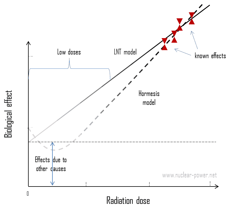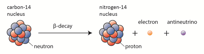
Facebook Instagram Youtube Twitter

What is Electron-hole Pair in Semiconductors – Definition

In the semiconductor, free charge carriers are electrons and electron holes (electron-hole pair). Electrons and holes are created by excitation of electron. Material Properties

extrinsic - doped semiconductor - p-type - acceptorIn the semiconductor, free charge carriers are electrons and electron holes (electron-hole pairs). Electrons and holes are created by excitation of electron from valence band to the conduction band. An electron hole (often simply called a hole) is the lack of an electron at a position where one could exist in an atom or atomic lattice. It is one of the two types of charge carriers that are responsible for creating electric current in semiconducting materials. Since in a normal atom or crystal lattice the negative charge of the electrons is balanced by the positive charge of the atomic nuclei, the absence of an electron leaves a net positive charge at the hole’s location. Positively charged holes can move from atom to atom in semiconducting materials as electrons leave their positions. When an electron meets with a hole, they recombine and these free carriers effectively vanish. The recombination means an electron which has been excited from the valence band to the conduction band falls back to the empty state in the valence band, known as the holes.

The conductivity of a semiconductor can be modeled in terms of the band theory of solids. The band model of a semiconductor suggests that at ordinary temperatures there is a finite possibility that electrons can reach the conduction band and contribute to electrical conduction. In the semiconductor, free charge carriers (electron-hole pairs) are created by excitation of electron from valence band to the conduction band. This excitation left a hole in the valence band which behaves as positive charge and an electron-hole pair is created. Holes can sometimes be confusing as they are not physical particles in the way that electrons are, rather they are the absence of an electron in an atom. Holes can move from atom to atom in semiconducting materials as electrons leave their positions.

Electron Excitation in Semiconductors

Energy for the excitation can be obtained by different ways.

Thermal Excitation

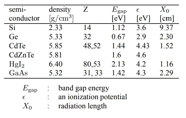
Electron-hole pairs are constantly generated from thermal energy as well, in the absence of any external energy source. Thermal excitation does not require any other form of starting impulse. This phenomenon occurs also at room temperature. It is caused by impurities, irregularity in structure lattice or by dopant. It strongly depends on the Egap (a distance between valence and conduction band), so that for lower Egap a number of thermally excited charge carriers increases. Since thermal excitation results in the detector noise, active cooling is required for some types of semiconductors (e.g. germanium). Detectors based on silicon have sufficiently low noise even by room temperature. This is caused by the large band gap of silicon (Egap= 1.12 eV), which allows us to operate the detector at room temperature, but cooling is prefered to reduce noise.

Optical Excitation

Note that, energy of a single photon of visible light spectrum is comparable with these band gaps. Photons of wave lengths 700 nm – 400 nm have energies of  1.77 eV 3.10 eV. As a result, also visible light is able to excite electrons to the conduction band. Actually, this is the principle of photovoltaic panels that generate electric current.

Excitation by Ionizing Radiation

Electrons may reach the conduction band, when they are excited by ionizing radiation (i.e. they must obtain energy higher than Egap). In general, heavy charged particles transfer energy mostly by:

  • Excitation. The charged particle can transfer energy to the atom, raising electrons to a higher energy levels.
  • Ionization. Ionization can occur, when the charged particle have enough energy to remove an electron. This results in a creation of ion pairs in surrounding matter.

A convenient variable that describes the ionization properties of surrounding medium is the stopping power. The classical expression that describes the specific energy loss is known as the Bethe formula. For alpha particles and heavier particles the stopping power of most materials is very high for heavy charged particles and these particles have very short ranges.

In addition to these interactions, beta particles also lose energy by radiative process known as the bremsstrahlung. From classical theory, when a charged particle is accelerated or decelerated, it must radiate energy and the deceleration radiation is known as the bremsstrahlung (“braking radiation”).

Photons (gamma rays and X-rays) can ionize atoms directly (despite they are electrically neutral) through the Photoelectric effect and the Compton effect, but secondary (indirect) ionization is much more significant. Although a large number of possible interactions are known, there are three key interaction mechanisms  with matter.

In all cases, a particle of ionizing radiation deposits a portion of its energy along its path. Particle passing through the detector ionizes the atoms of semiconductor, producing the electron-hole pairs. For example, typical thickness of silicon detector is about 300 µm so the number of generated electron-hole pairs by minimum ionizing particle (MIP) passing perpendicular through the detector is about 3.2 x 10⁴. This value is minor in comparison the total number of free carriers in intrinsic semiconductor of a surface of 1 cm2 and the same thickness. Note that, a sample of pure germanium at 20 °C contains about 1.26×10²¹ atoms, but also contains 7.5 x 10¹¹ free electrons and 7.5 x 10¹¹ holes constantly generated from thermodynamics/what-is-energy-physics/internal-energy-thermal-energy/”>thermal energy. As can be seen, the signal to noise ratio (S/N) would be minimal. The addition of 0.001% of arsenic (an impurity) donates an extra 1015 free electrons in the same volume and the electrical conductivity is increased by a factor of 10,000. In doped material the signal to noise ratio (S/N) would be even smaller. Cooling of the semiconductor is one way to lower this ratio.

Improvement can be reached by use of a reverse-bias voltage to the P-N junction to deplete the detector of free carriers, which is the principle of the most silicon radiation detectors. In this case, negative voltage is applied to the p-side and positive to the second one. Holes in the p-region are attracted from the junction towards the p contact and similarly for electrons and the n contact.

References:

Radiation Protection:

  1. Knoll, Glenn F., Radiation Detection and Measurement 4th Edition, Wiley, 8/2010. ISBN-13: 978-0470131480.
  2. Stabin, Michael G., Radiation Protection and Dosimetry: An Introduction to Health Physics, Springer, 10/2010. ISBN-13: 978-1441923912.
  3. Martin, James E., Physics for Radiation Protection 3rd Edition, Wiley-VCH, 4/2013. ISBN-13: 978-3527411764.
  4. U.S.NRC, NUCLEAR REACTOR CONCEPTS
  5. U.S. Department of Energy, Instrumantation and Control. DOE Fundamentals Handbook, Volume 2 of 2. June 1992.

Nuclear and Reactor Physics:

  1. J. R. Lamarsh, Introduction to Nuclear Reactor Theory, 2nd ed., Addison-Wesley, Reading, MA (1983).
  2. J. R. Lamarsh, A. J. Baratta, Introduction to Nuclear Engineering, 3d ed., Prentice-Hall, 2001, ISBN: 0-201-82498-1.
  3. W. M. Stacey, Nuclear Reactor Physics, John Wiley & Sons, 2001, ISBN: 0- 471-39127-1.
  4. Glasstone, Sesonske. Nuclear Reactor Engineering: Reactor Systems Engineering, Springer; 4th edition, 1994, ISBN: 978-0412985317
  5. W.S.C. Williams. Nuclear and Particle Physics. Clarendon Press; 1 edition, 1991, ISBN: 978-0198520467
  6. G.R.Keepin. Physics of Nuclear Kinetics. Addison-Wesley Pub. Co; 1st edition, 1965
  7. Robert Reed Burn, Introduction to Nuclear Reactor Operation, 1988.
  8. U.S. Department of Energy, Nuclear Physics and Reactor Theory. DOE Fundamentals Handbook, Volume 1 and 2. January 1993.
  9. Paul Reuss, Neutron Physics. EDP Sciences, 2008. ISBN: 978-2759800414.

See also:

Properties of Semiconductors

We hope, this article, Electron-hole Pair in Semiconductors, helps you. If so, give us a like in the sidebar. Main purpose of this website is to help the public to learn some interesting and important information about materials and their properties.

What is Fermi Level in Semiconductors – Definition

The Fermi level is the surface of Fermi sea at absolute zero where no electrons will have enough energy to rise above the surface. Fermi Level in Semiconductors

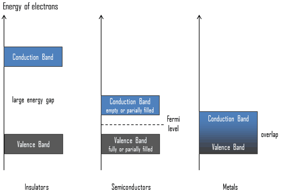
Valence Band - Conduction Band - Band GapIn solid-state physics, the valence band and conduction band are the bands closest to the Fermi level and thus determine the electrical conductivity of the solid. In electrical insulators and semiconductors, the valence band is the highest range of electron energies in which electrons are normally present at absolute zero temperature. For example, a silicon atom has fourteen electrons. In the ground state, they are arranged in the electron configuration [Ne]3s23p2. Of these, four are valence electrons, occupying the 3s orbital and two of the 3p orbitals. The distinction between the valence and conduction bands is meaningless in metals, because conduction occurs in one or more partially filled bands that take on the properties of both the valence and conduction bands.

Fermi Level

The term “Fermi level” comes from Fermi-Dirac statistics, which describes a distribution of particles over energy states in systems consisting of fermions (electrons) that obey the Pauli exclusion principle. Since they cannot exist in identical energy states, Fermi level is the term used to describe the top of the collection of electron energy levels at thermodynamics/thermodynamic-properties/what-is-temperature-physics/absolute-zero-temperature/”>absolute zero temperature. The Fermi level is the surface of Fermi sea at absolute zero where no electrons will have enough energy to rise above the surface. In metals, the Fermi level lies in the hypothetical conduction band giving rise to free conduction electrons. In semiconductors the position of the Fermi level is within the band gap, approximately in the middle of the band gap.

References:

Radiation Protection:

  1. Knoll, Glenn F., Radiation Detection and Measurement 4th Edition, Wiley, 8/2010. ISBN-13: 978-0470131480.
  2. Stabin, Michael G., Radiation Protection and Dosimetry: An Introduction to Health Physics, Springer, 10/2010. ISBN-13: 978-1441923912.
  3. Martin, James E., Physics for Radiation Protection 3rd Edition, Wiley-VCH, 4/2013. ISBN-13: 978-3527411764.
  4. U.S.NRC, NUCLEAR REACTOR CONCEPTS
  5. U.S. Department of Energy, Instrumantation and Control. DOE Fundamentals Handbook, Volume 2 of 2. June 1992.

Nuclear and Reactor Physics:

  1. J. R. Lamarsh, Introduction to Nuclear Reactor Theory, 2nd ed., Addison-Wesley, Reading, MA (1983).
  2. J. R. Lamarsh, A. J. Baratta, Introduction to Nuclear Engineering, 3d ed., Prentice-Hall, 2001, ISBN: 0-201-82498-1.
  3. W. M. Stacey, Nuclear Reactor Physics, John Wiley & Sons, 2001, ISBN: 0- 471-39127-1.
  4. Glasstone, Sesonske. Nuclear Reactor Engineering: Reactor Systems Engineering, Springer; 4th edition, 1994, ISBN: 978-0412985317
  5. W.S.C. Williams. Nuclear and Particle Physics. Clarendon Press; 1 edition, 1991, ISBN: 978-0198520467
  6. G.R.Keepin. Physics of Nuclear Kinetics. Addison-Wesley Pub. Co; 1st edition, 1965
  7. Robert Reed Burn, Introduction to Nuclear Reactor Operation, 1988.
  8. U.S. Department of Energy, Nuclear Physics and Reactor Theory. DOE Fundamentals Handbook, Volume 1 and 2. January 1993.
  9. Paul Reuss, Neutron Physics. EDP Sciences, 2008. ISBN: 978-2759800414.

See also:

Properties of Semiconductors

We hope, this article, Fermi Level in Semiconductors, helps you. If so, give us a like in the sidebar. Main purpose of this website is to help the public to learn some interesting and important information about materials and their properties.

What is Band Gap – Energy Gap in Semiconductors – Definition

In solid-state physics, this energy gap or band gap is an energy range between valence band and conduction band where electron states are forbidden. Material Properties

semiconductor detectors - table of parametersThe name semiconductor comes from the fact that these materials  have an electrical conductivity between that of a metal, like copper, gold, etc. and an insulator, such as glass. They have an energy gap less than 4eV (about 1eV). In solid-state physics, this energy gap or band gap is an energy range between valence band and conduction band where electron states are forbidden. Properties of semiconductors are determined by the energy gap between valence and conduction bands. To understand, what is semiconductor, we have to define these terms.

In solid-state physics, the energy gap or the band gap is an energy range between valence band and conduction band where electron states are forbidden. In contrast to conductors, electrons in a semiconductor must obtain energy (e.g. from ionizing radiation) to cross the band gap and to reach the conduction band. Band gaps are naturally different for different materials. For example, diamond is a wide-band gap semiconductor (Egap = 5.47 eV) with high potential as an electronic device material in many devices. On the other side, germanium has a small band gap energy (Egap = 0.67 eV), which requires to operate the detector at cryogenic temperatures.

References:

Radiation Protection:

  1. Knoll, Glenn F., Radiation Detection and Measurement 4th Edition, Wiley, 8/2010. ISBN-13: 978-0470131480.
  2. Stabin, Michael G., Radiation Protection and Dosimetry: An Introduction to Health Physics, Springer, 10/2010. ISBN-13: 978-1441923912.
  3. Martin, James E., Physics for Radiation Protection 3rd Edition, Wiley-VCH, 4/2013. ISBN-13: 978-3527411764.
  4. U.S.NRC, NUCLEAR REACTOR CONCEPTS
  5. U.S. Department of Energy, Instrumantation and Control. DOE Fundamentals Handbook, Volume 2 of 2. June 1992.

Nuclear and Reactor Physics:

  1. J. R. Lamarsh, Introduction to Nuclear Reactor Theory, 2nd ed., Addison-Wesley, Reading, MA (1983).
  2. J. R. Lamarsh, A. J. Baratta, Introduction to Nuclear Engineering, 3d ed., Prentice-Hall, 2001, ISBN: 0-201-82498-1.
  3. W. M. Stacey, Nuclear Reactor Physics, John Wiley & Sons, 2001, ISBN: 0- 471-39127-1.
  4. Glasstone, Sesonske. Nuclear Reactor Engineering: Reactor Systems Engineering, Springer; 4th edition, 1994, ISBN: 978-0412985317
  5. W.S.C. Williams. Nuclear and Particle Physics. Clarendon Press; 1 edition, 1991, ISBN: 978-0198520467
  6. G.R.Keepin. Physics of Nuclear Kinetics. Addison-Wesley Pub. Co; 1st edition, 1965
  7. Robert Reed Burn, Introduction to Nuclear Reactor Operation, 1988.
  8. U.S. Department of Energy, Nuclear Physics and Reactor Theory. DOE Fundamentals Handbook, Volume 1 and 2. January 1993.
  9. Paul Reuss, Neutron Physics. EDP Sciences, 2008. ISBN: 978-2759800414.

See also:

Properties of Semiconductors

We hope, this article, Band Gap – Energy Gap in Semiconductors, helps you. If so, give us a like in the sidebar. Main purpose of this website is to help the public to learn some interesting and important information about materials and their properties.

What is Semiconductivity – Band Theory – Definition

Semiconductivity is determined by the energy gap between valence and conduction bands. To understand, what is semiconductor, we have to define these terms. Material Properties

In general, semiconductors are materials, inorganic or organic, which have the ability to control their conduction depending on chemical structure, temperature, illumination, and presence of dopants. The name semiconductor comes from the fact that these materials  have an electrical conductivity between that of a metal, like copper, gold, etc. and an insulator, such as glass. They have an energy gap less than 4eV (about 1eV). In solid-state physics, this energy gap or band gap is an energy range between valence band and conduction band where electron states are forbidden. In contrast to conductors, electrons in a semiconductor must obtain energy (e.g. from ionizing radiation) to cross the band gap and to reach the conduction band. Properties of semiconductors are determined by the energy gap between valence and conduction bands. To understand, what is semiconductor, we have to define these terms.

Properties of Semiconductors

To understand the difference between metals, semiconductors and electrical insulators, we have to define the following terms from solid-state physics:

  • Valence Band - Conduction Band - Band GapValence Band. In solid-state physics, the valence band and conduction band are the bands closest to the Fermi level and thus determine the electrical conductivity of the solid. In electrical insulators and semiconductors, the valence band is the highest range of electron energies in which electrons are normally present at absolute zero temperature. For example, a silicon atom has fourteen electrons. In the ground state, they are arranged in the electron configuration [Ne]3s23p2. Of these, four are valence electrons, occupying the 3s orbital and two of the 3p orbitals. The distinction between the valence and conduction bands is meaningless in metals, because conduction occurs in one or more partially filled bands that take on the properties of both the valence and conduction bands.
  • Conduction Band. In solid-state physics, the valence band and conduction band are the bands closest to the Fermi level and thus determine the electrical conductivity of the solid. In electrical insulators and semiconductors, the conduction band is the lowest range of vacant electronic states. On a graph of the electronic band structure of a material, the valence band is located below the Fermi level, while the conduction band is located above it. In semiconductors, electrons may reach the conduction band, when they are excited, for example, by ionizing radiation (i.e. they must obtain energy higher than Egap). For example, diamond is a wide-band gap semiconductor (Egap = 5.47 eV) with high potential as an electronic device material in many devices. On the other side, germanium has a small band gap energy (Egap = 0.67 eV), which requires to operate the detector at cryogenic temperatures. The distinction between the valence and conduction bands is meaningless in metals, because conduction occurs in one or more partially filled bands that take on the properties of both the valence and conduction bands.
  • Band Gap. In solid-state physics, the energy gap or the band gap is an energy range between valence band and conduction band where electron states are forbidden. In contrast to conductors, electrons in a semiconductor must obtain energy (e.g. from ionizing radiation) to cross the band gap and to reach the conduction band. Band gaps are naturally different for different materials. For example, diamond is a wide-band gap semiconductor (Egap = 5.47 eV) with high potential as an electronic device material in many devices. On the other side, germanium has a small band gap energy (Egap = 0.67 eV), which requires to operate the detector at cryogenic temperatures.
  • Fermi Level. The term “Fermi level” comes from Fermi-Dirac statistics, which describes a distribution of particles over energy states in systems consisting of fermions (electrons) that obey the Pauli exclusion principle. Since they cannot exist in identical energy states, Fermi level is the term used to describe the top of the collection of electron energy levels at thermodynamics/thermodynamic-properties/what-is-temperature-physics/absolute-zero-temperature/”>absolute zero temperature. The Fermi level is the surface of Fermi sea at absolute zero where no electrons will have enough energy to rise above the surface. In metals, the Fermi level lies in the hypothetical conduction band giving rise to free conduction electrons. In semiconductors the position of the Fermi level is within the band gap, approximately in the middle of the band gap.
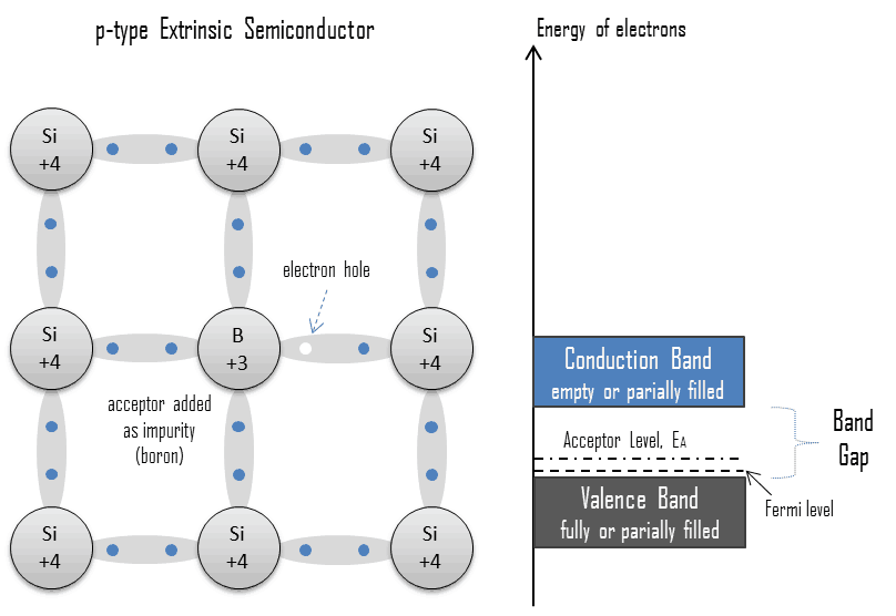
  • extrinsic - doped semiconductor - p-type - acceptorElectron-hole Pair. In the semiconductor, free charge carriers are electrons and electron holes (electron-hole pairs). Electrons and holes are created by excitation of electron from valence band to the conduction band. An electron hole (often simply called a hole) is the lack of an electron at a position where one could exist in an atom or atomic lattice. It is one of the two types of charge carriers that are responsible for creating electric current in semiconducting materials. Since in a normal atom or crystal lattice the negative charge of the electrons is balanced by the positive charge of the atomic nuclei, the absence of an electron leaves a net positive charge at the hole’s location. Positively charged holes can move from atom to atom in semiconducting materials as electrons leave their positions. When an electron meets with a hole, they recombine and these free carriers effectively vanish. The recombination means an electron which has been excited from the valence band to the conduction band falls back to the empty state in the valence band, known as the holes.

The conductivity of a semiconductor can be modeled in terms of the band theory of solids. The band model of a semiconductor suggests that at ordinary temperatures there is a finite possibility that electrons can reach the conduction band and contribute to electrical conduction. In the semiconductor, free charge carriers (electron-hole pairs) are created by excitation of electron from valence band to the conduction band. This excitation left a hole in the valence band which behaves as positive charge and an electron-hole pair is created. Holes can sometimes be confusing as they are not physical particles in the way that electrons are, rather they are the absence of an electron in an atom. Holes can move from atom to atom in semiconducting materials as electrons leave their positions.

References:

Radiation Protection:

  1. Knoll, Glenn F., Radiation Detection and Measurement 4th Edition, Wiley, 8/2010. ISBN-13: 978-0470131480.
  2. Stabin, Michael G., Radiation Protection and Dosimetry: An Introduction to Health Physics, Springer, 10/2010. ISBN-13: 978-1441923912.
  3. Martin, James E., Physics for Radiation Protection 3rd Edition, Wiley-VCH, 4/2013. ISBN-13: 978-3527411764.
  4. U.S.NRC, NUCLEAR REACTOR CONCEPTS
  5. U.S. Department of Energy, Instrumantation and Control. DOE Fundamentals Handbook, Volume 2 of 2. June 1992.

Nuclear and Reactor Physics:

  1. J. R. Lamarsh, Introduction to Nuclear Reactor Theory, 2nd ed., Addison-Wesley, Reading, MA (1983).
  2. J. R. Lamarsh, A. J. Baratta, Introduction to Nuclear Engineering, 3d ed., Prentice-Hall, 2001, ISBN: 0-201-82498-1.
  3. W. M. Stacey, Nuclear Reactor Physics, John Wiley & Sons, 2001, ISBN: 0- 471-39127-1.
  4. Glasstone, Sesonske. Nuclear Reactor Engineering: Reactor Systems Engineering, Springer; 4th edition, 1994, ISBN: 978-0412985317
  5. W.S.C. Williams. Nuclear and Particle Physics. Clarendon Press; 1 edition, 1991, ISBN: 978-0198520467
  6. G.R.Keepin. Physics of Nuclear Kinetics. Addison-Wesley Pub. Co; 1st edition, 1965
  7. Robert Reed Burn, Introduction to Nuclear Reactor Operation, 1988.
  8. U.S. Department of Energy, Nuclear Physics and Reactor Theory. DOE Fundamentals Handbook, Volume 1 and 2. January 1993.
  9. Paul Reuss, Neutron Physics. EDP Sciences, 2008. ISBN: 978-2759800414.

See also:

Properties of Semiconductors

We hope, this article, Semiconductivity – Band Theory, helps you. If so, give us a like in the sidebar. Main purpose of this website is to help the public to learn some interesting and important information about materials and their properties.

What is Conduction and Valence Band in Semiconductors – Definition

In solid-state physics, the valence band and conduction band are the bands closest to the Fermi level and thus determine the electrical conductivity of the semiconductor. Material Properties

To understand the difference between metals, semiconductors and electrical insulators, we have to define the following terms from solid-state physics:

Valence Band - Conduction Band - Band GapValence Band

In solid-state physics, the valence band and conduction band are the bands closest to the Fermi level and thus determine the electrical conductivity of the solid. In electrical insulators and semiconductors, the valence band is the highest range of electron energies in which electrons are normally present at absolute zero temperature. For example, a silicon atom has fourteen electrons. In the ground state, they are arranged in the electron configuration [Ne]3s23p2. Of these, four are valence electrons, occupying the 3s orbital and two of the 3p orbitals. The distinction between the valence and conduction bands is meaningless in metals, because conduction occurs in one or more partially filled bands that take on the properties of both the valence and conduction bands.

Conduction Band

In solid-state physics, the valence band and conduction band are the bands closest to the Fermi level and thus determine the electrical conductivity of the solid. In electrical insulators and semiconductors, the conduction band is the lowest range of vacant electronic states. On a graph of the electronic band structure of a material, the valence band is located below the Fermi level, while the conduction band is located above it. In semiconductors, electrons may reach the conduction band, when they are excited, for example, by ionizing radiation (i.e. they must obtain energy higher than Egap). For example, diamond is a wide-band gap semiconductor (Egap = 5.47 eV) with high potential as an electronic device material in many devices. On the other side, germanium has a small band gap energy (Egap = 0.67 eV), which requires to operate the detector at cryogenic temperatures. The distinction between the valence and conduction bands is meaningless in metals, because conduction occurs in one or more partially filled bands that take on the properties of both the valence and conduction bands.

References:

Radiation Protection:

  1. Knoll, Glenn F., Radiation Detection and Measurement 4th Edition, Wiley, 8/2010. ISBN-13: 978-0470131480.
  2. Stabin, Michael G., Radiation Protection and Dosimetry: An Introduction to Health Physics, Springer, 10/2010. ISBN-13: 978-1441923912.
  3. Martin, James E., Physics for Radiation Protection 3rd Edition, Wiley-VCH, 4/2013. ISBN-13: 978-3527411764.
  4. U.S.NRC, NUCLEAR REACTOR CONCEPTS
  5. U.S. Department of Energy, Instrumantation and Control. DOE Fundamentals Handbook, Volume 2 of 2. June 1992.

Nuclear and Reactor Physics:

  1. J. R. Lamarsh, Introduction to Nuclear Reactor Theory, 2nd ed., Addison-Wesley, Reading, MA (1983).
  2. J. R. Lamarsh, A. J. Baratta, Introduction to Nuclear Engineering, 3d ed., Prentice-Hall, 2001, ISBN: 0-201-82498-1.
  3. W. M. Stacey, Nuclear Reactor Physics, John Wiley & Sons, 2001, ISBN: 0- 471-39127-1.
  4. Glasstone, Sesonske. Nuclear Reactor Engineering: Reactor Systems Engineering, Springer; 4th edition, 1994, ISBN: 978-0412985317
  5. W.S.C. Williams. Nuclear and Particle Physics. Clarendon Press; 1 edition, 1991, ISBN: 978-0198520467
  6. G.R.Keepin. Physics of Nuclear Kinetics. Addison-Wesley Pub. Co; 1st edition, 1965
  7. Robert Reed Burn, Introduction to Nuclear Reactor Operation, 1988.
  8. U.S. Department of Energy, Nuclear Physics and Reactor Theory. DOE Fundamentals Handbook, Volume 1 and 2. January 1993.
  9. Paul Reuss, Neutron Physics. EDP Sciences, 2008. ISBN: 978-2759800414.

See also:

Properties of Semiconductors

We hope, this article, Conduction and Valence Band in Semiconductors, helps you. If so, give us a like in the sidebar. Main purpose of this website is to help the public to learn some interesting and important information about materials and their properties.

What is Ionization – Definition

Ionization is the process in which an atom or a molecule gains or loses electrons to form charged ion. Ionization can result from the loss of an electron after collisions with energetic subatomic particles. Material Properties

What is Ionization

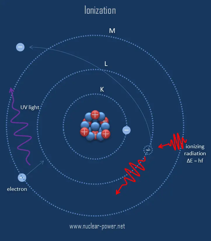
Ionization - DefinitionIonization is the process in which an atom or a molecule gains or loses electrons to form charged ion. Ionization can result from the loss of an electron after collisions with energetic subatomic particles, collisions with other atoms, molecules and ions, or through the interaction with electromagnetic radiation. In general, ionizing radiation is any radiation (particles or electromagnetic waves) that carries enough energy to knock electrons from atoms or molecules, thereby ionizing them. For ionizing radiation, the kinetic energy of particles (photons, electrons, etc.) is sufficient and the particle can ionize (to form ion by losing electrons) target atoms to form ions.

The boundary between ionizing and non-ionizing radiation is not sharply defined, since different molecules and atoms ionize at different energies. Gamma rays, X-rays, and the higher ultraviolet part of the spectrum are ionizing, whereas the lower ultraviolet, visible light (including laser light), infrared, microwaves, and radio waves are considered non-ionizing radiation.

Ionization Energy

Ionization energy, also called ionization potential, is the energy necessary to remove an electron from the neutral atom.

X + energy → X+ + e−

where X is any atom or molecule capable of being ionized, X+ is that atom or molecule with an electron removed (positive ion), and e− is the removed electron.

A nitrogen atom, for example, requires the following ionization energy to remove the outermost electron.

N + IE → N+ + e−        IE = 14.5 eV

The ionization energy associated with removal of the first electron is most commonly used. The nth ionization energy refers to the amount of energy required to remove an electron from the species with a charge of (n-1).

1st ionization energy

X → X+ + e−

2nd ionization energy

X+ → X2+ + e−

3rd ionization energy

X2+ → X3+ + e−

Ionization Energy for different Elements

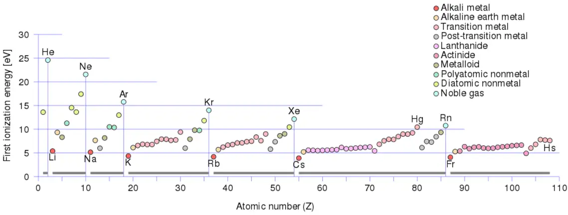
There is an ionization energy for each successive electron removed. The electrons that circle the nucleus move in fairly well-defined orbits. Some of these electrons are more tightly bound in the atom than others. For example, only 7.38 eV is required to remove the outermost electron from a lead atom, while 88,000 eV is required to remove the innermost electron. Helps to understand reactivity of elements (especially metals, which lose electrons).

In general, the ionization energy increases moving up a group and moving left to right across a period. Moreover:

  • Ionization energy is lowest for the alkali metals which have a single electron outside a closed shell.
  • Ionization energy increases across a row on the periodic maximum for the noble gases which have closed shells

For example, sodium requires only 496 kJ/mol or 5.14 eV/atom to ionize it. On the other hand neon, the noble gas, immediately preceding it in the periodic table, requires 2081 kJ/mol or 21.56 eV/atom.

Ionization energy
Source: wikipedia.org License: CC BY-SA 3.0
References:

Radiation Protection:

  1. Knoll, Glenn F., Radiation Detection and Measurement 4th Edition, Wiley, 8/2010. ISBN-13: 978-0470131480.
  2. Stabin, Michael G., Radiation Protection and Dosimetry: An Introduction to Health Physics, Springer, 10/2010. ISBN-13: 978-1441923912.
  3. Martin, James E., Physics for Radiation Protection 3rd Edition, Wiley-VCH, 4/2013. ISBN-13: 978-3527411764.
  4. U.S.NRC, NUCLEAR REACTOR CONCEPTS
  5. U.S. Department of Energy, Instrumantation and Control. DOE Fundamentals Handbook, Volume 2 of 2. June 1992.

Nuclear and Reactor Physics:

  1. J. R. Lamarsh, Introduction to Nuclear Reactor Theory, 2nd ed., Addison-Wesley, Reading, MA (1983).
  2. J. R. Lamarsh, A. J. Baratta, Introduction to Nuclear Engineering, 3d ed., Prentice-Hall, 2001, ISBN: 0-201-82498-1.
  3. W. M. Stacey, Nuclear Reactor Physics, John Wiley & Sons, 2001, ISBN: 0- 471-39127-1.
  4. Glasstone, Sesonske. Nuclear Reactor Engineering: Reactor Systems Engineering, Springer; 4th edition, 1994, ISBN: 978-0412985317
  5. W.S.C. Williams. Nuclear and Particle Physics. Clarendon Press; 1 edition, 1991, ISBN: 978-0198520467
  6. G.R.Keepin. Physics of Nuclear Kinetics. Addison-Wesley Pub. Co; 1st edition, 1965
  7. Robert Reed Burn, Introduction to Nuclear Reactor Operation, 1988.
  8. U.S. Department of Energy, Nuclear Physics and Reactor Theory. DOE Fundamentals Handbook, Volume 1 and 2. January 1993.
  9. Paul Reuss, Neutron Physics. EDP Sciences, 2008. ISBN: 978-2759800414.

See also:

Radiation Detection

We hope, this article, Ionization, helps you. If so, give us a like in the sidebar. Main purpose of this website is to help the public to learn some interesting and important information about materials and their properties.

What is Radioactive Contamination – Definition

Radioactive contamination is referred to as the presence of an unwanted radioactive substances on surfaces, or within solids (including the human body), liquids or gases, where their presence is unintended or undesirable. Material Properties
radioactive contamination
Radioactive contamination consist of radioactive material, that generate ionizing radiation. It is the source of radiation, not radiation itself.

Contamination is generally referred to as the presence of an unwanted constituent, harmful substance or impurity in a location (material, physical body, natural environment, workplace) where it is not intended or desired to be. Contamination has much more general meaning, since is can be defined in disciplines such as chemistry, environmental protection, radiation protection or agriculture.

Radioactive contamination is referred to as the presence of an unwanted radioactive substances on surfaces, or within solids (including the human body), liquids or gases, where their presence is unintended or undesirable. Radioactive contamination consist of radioactive atoms (material) that have escaped the system or structure that would normally contain them. Since radioactive contamination is radioactive material, ionizing radiation is emitted by the contamination. It is very important, which material (which radioisotope) is the radioactive contaminant. It is also very important to distinguish between radioactive contamination and radiation itself.

Contamination versus Radiation

Radioactive contamination consist of radioactive material, that generate ionizing radiation. It is the source of radiation, not radiation itself. Anytime that radioactive material is not in a sealed radioactive source container and might be spread onto other objects, radioactive contamination is a possibility. Radioactive contamination may be characterized by following points:

  • Radioactive contamination consist of radioactive material (contaminants), that may be solid, liquid or gaseous. Large contaminants can be even visible, but you cannot see radiation produced.
  • When released, contaminants can be spread by air, water or just by mechanical contact.
  • We cannot shield contamination.
  • We can mitigate contamination by protecting integrity of barriers (source container, fuel cladding, reactor vessel, containment building)
  • Since contaminants interact chemically, they may be contained within objects such as the human body.
  • We can rid of contamination by many mechanical, chemical (decontaminate surfaces), or biological processes (biological half-life).
  • It is of the highest importance, which material is the radioactive contaminant (half-life, mode of decay, energy).

Ionizing radiation is formed by high-energy particles (photons, electrons, etc.), that can penetrate matter and ionize (to form ion by losing electrons) target atoms to form ions. Radiation exposure is the consequence of the presence nearby the source of radiation. Radiation exposure as a quantity is defined as a measure of the ionization of material due to ionizing radiation. The danger of ionizing radiation lies in the fact that the radiation is invisible and not directly detectable by human senses. People can neither see nor feel radiation, yet it deposits energy to the molecules of the body. The energy is transferred in small quantities for each interaction between radiation and a molecule and there are usually many such interactions. Unlike radioactive contamination, radiation may be characterized by following points:

  • Radiation consist consist of high-energy particles that can penetrate matter and ionize (to form ion by losing electrons) target atoms. Radiation is invisible, and not directly detectable by human senses. It must be noted, beta radiation is indirectly visible due to cherenkov radiation.
  • Unlike contamination, radiation cannot be spread by any medium. It travels through materials until it loses its energy. We can shield radiation (e.g. by standing around the corner).
  • Exposure to ionizing does not necessary mean, that the object becomes radioactive (except very rare neutron radiation).
  • Radiation can penetrate barriers, but sufficiently thick barrier can minimize all effects.
  • Unlike contaminants, radiation cannot interact chemically with matter and cannot be bound inside body.
  • It is not important, which material is the source of certain radiation. Only type of radiation and energy matters.

There is one common feature, natural radiation and natural contaminants are all around us. In, around, and above the world we live in. It is a natural energy force that surrounds us. It is a part of our natural world that has been here since the birth of our planet. All living creatures, from the beginning of time, have been, and are still being, exposed to ionizing radiation. Natural background radiation is ionizing radiation, that originates from a variety of natural sources. All living creatures, from the beginning of time, have been, and are still being, exposed to ionizing radiation. This radiation is not associated with any human activity.  There are radioactive isotopes in our bodies, houses, air, water and in the soil. We all are also exposed to radiation from outer space.

Types of Contamination

Radioactive materials may exist on surfaces or in volumes of material or air, and specialist techniques are used to measure the levels of contamination by detection of the emitted radiation. We can distinguish between following types of contamination:

Surface Contamination

Surface contamination means that radioactive material has been deposited on surfaces (such as walls, floors). It may be loosely deposited, much like ordinary dust, or it may be quite firmly fixed by chemical reaction. This distinction is important, and we classify surface contamination on the basis of how easily it can be removed:

  • Free Contamination. In the case of free contamination (or loose contamination), the radioactive material can be spread. This is surface contamination that can easily be removed with simple decontamination methods. For example, if dust particles containing various radioisotopes land on the person’s skin or garments, we can clean it up or remove clothes. Once a person has been decontaminated, all of the particulate radioactivity sources are eliminated, and the individual is no longer contaminated. Free contamination is also a more serious hazard than fixed contamination, because dust particles can become airborne and they can be easily ingested. This leads to an internal exposure by radioactive contaminants. Although almost all contaminants are beta radioactive with accompanying gamma emission, but there is also the possibility of alpha contamination in any nuclear fuel handling areas.
  • Fixed Contamination. In the case of fixed contamination, the radioactive material cannot be spread, since it is chemically or mechanically bound to structures. It cannot be removed by normal cleaning methods. Fixed contamination is a less serious hazard than free contamination, it cannot be re-suspended or transferred to skin. Therefore the hazard is usually an external one only. On the other hand, it depends on the level of contamination. Although almost all contaminants are beta radioactive with accompanying gamma emission, but there is also the possibility of alpha contamination in any nuclear fuel handling areas. Unless the level of contamination is very severe, the gamma radiation dose rate will be small and external exposure will be significant only in contact with, or very close to, the contaminated surfaces. Since beta particles are less penetrating than gamma rays, the beta dose rate can be high only at contact. A value of 1 mSv/h at contact for a contamination level of 400 – 500 Bq/cm2 is fairly representative.
Calculation of Shielded Dose Rate in Sieverts from Contaminated Surface

Assume a surface, which is contamined by 1.0 Ci of 137Cs. Assume that this contaminant can be aproximated by the point isotropic source which contains 1.0 Ci of 137Cs, which has a half-life of 30.2 years. Note that the relationship between half-life and the amount of a radionuclide required to give an activity of one curie is shown below. This amount of material can be calculated using λ, which is the decay constant of certain nuclide:

Curie - Unit of Activity

About 94.6 percent decays by beta emission to a metastable nuclear isomer of barium: barium-137m. The main photon peak of Ba-137m is 662 keV. For this calculation, assume that all decays go through this channel.

Calculate the primary photon dose rate, in sieverts per hour (Sv.h-1), at the outer surface of a 5 cm thick lead shield. Then calculate the equivalent and effective dose rates for two cases.

  1. Assume that this external radiation field penetrates uniformly through the whole body. That means: Calculate the effective whole-body dose rate.
  2. Assume that this external radiation field penetrates only lungs and the other organs are completely shielded. That means: Calculate the effective dose rate.

Note that, primary photon dose rate neglects all secondary particles. Assume that the effective distance of the source from the dose point is 10 cm. We shall also assume that the dose point is soft tissue and it can reasonably be simulated by water and we use the mass energy absorption coefficient for water.

See also: Gamma Ray Attenuation

See also: Shielding of Gamma Rays

Solution:

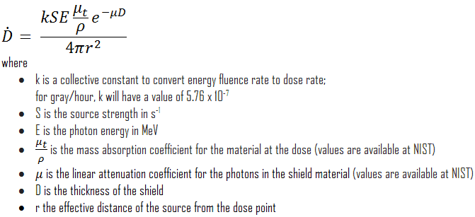
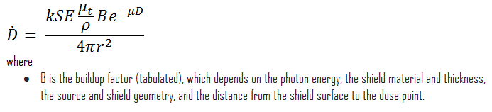
The primary photon dose rate is attenuated exponentially, and the dose rate from primary photons, taking account of the shield, is given by:

dose rate calculation

As can be seen, we do not account for the buildup of secondary radiation. If secondary particles are produced or if the primary radiation changes its energy or direction, then the effective attenuation will be much less.  This assumption generally underestimates the true dose rate, especially for thick shields and when the dose point is close to the shield surface, but this assumption simplifies all calculations. For this case the true dose rate (with the buildup of secondary radiation) will be more than two times higher.

To calculate the absorbed dose rate, we have to use in the formula:

  • k = 5.76 x 10⁻⁷
  • S = 3.7 x 10¹⁰ s-1
  • E = 0.662 MeV
  • μt/ρ =  0.0326 cm2/g (values are available at NIST)
  • μ =  1.289 cm-1 (values are available at NIST)
  • D = 5 cm
  • r = 10 cm

Result:

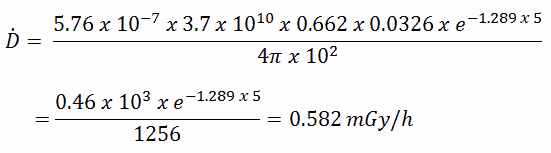
The resulting absorbed dose rate in grays per hour is then:

absorbed dose rate - gray - calculation

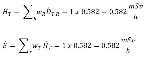
1) Uniform irradiation

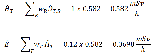
Since the radiation weighting factor for gamma rays is equal to one and we have assumed the uniform radiation field (the tissue weighting factor is also equal to unity), we can directly calculate the equivalent dose rate and the effective dose rate (E = HT) from the absorbed dose rate as:

calculation - effective dose - uniform

2) Partial irradiation

In this case we assume a partial irradiation of lungs only. Thus, we have to use the tissue weighting factor, which is equal to wT = 0.12. The radiation weighting factor for gamma rays is equal to one. As a result, we can calculate the effective dose rate as:

calculation - effective dose - non-uniform

Note that, if one part of the body (e.g.,the lungs) receives a radiation dose, it represents a risk for a particularly damaging effect (e.g., lung cancer). If the same dose is given to another organ it represents a different risk factor.

If we want to account for the buildup of secondary radiation, then we have to include the buildup factor. The extended formula for the dose rate is then:

absorbed dose rate - gray


——–

Airborne Contamination

This type of contamination is of particular importance in nuclear power plants, where it must be monitored. Contaminants can become airborne especially during reactor top head remove, reactor refueling, and during manipulations within spent fuel pool. The air can be contaminated with radioactive isotopes especially in particulate form, which poses a particular inhalation hazard. This contamination consists of various fission and activation products that enter the air in gaseous, vapour or particulate form. There are four types of airborne contamination in nuclear power plants, namely:

  • Particulates. Particulate activity is an internal hazard, because it can be inhaled. Transportable particulate material taken into the respiratory system will enter the blood stream and be carried to all parts of the body. Non-transportable particulates will stay in the lungs with a certain biological half-life. For example, Sr-90, Ra-226 and Pu-239 are radionuclides known as bone-seeking radionuclides. These radionuclides have long biological half-lives and are serious internal hazards. Once deposited in bone, they remain there essentially unchanged in amount during the lifetime of the individual. The continued action of the emitted alpha particles can cause significant injury: over many years they deposit all their energy in a tiny volume of tissue, because the range of the alpha particles is very short.
  • Noble gases. Radioactive noble gases, such as xenon-133, xenon-135 and  krypton-85 are present in reactor coolant especially when fuel leakages are present. As they appear in coolant, they become airborne and they can be inhaled. They are exhaled right after they are inhaled, because the body does not react chemically with them. If workers are working in a noble gas cloud, the external dose they will receive is about 1000 times greater than the internal dose. Because of this, we are only concerned about the external beta and gamma dose rates.
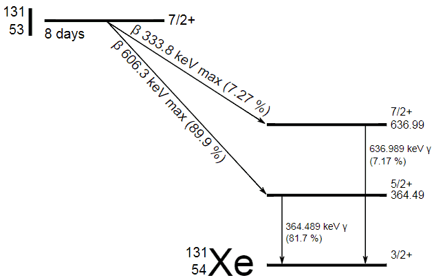
  • Iodine 131 - decay schemeRadioiodine. Radioiodine, iodine-131, is an important radioisotope of iodine. Radioiodine plays a major role as a radioactive isotope present in nuclear fission products, and it is a major contributor to the health hazards when released into the atmosphere during an accident. Iodine-131 has a half-life of 8.02 days. The target tissue for radioiodine exposure is the thyroid gland. The external beta and gamma dose from radioiodine present in the air is quite negligible when compared to the committed dose to the thyroid that would result from breathing this air. The biological half-life for iodine inside the human body is about 80 days (according to ICRP). Iodine in food is absorbed by the body and preferentially concentrated in the thyroid where it is needed for the functioning of that gland. When 131I is present in high levels in the environment from radioactive fallout, it can be absorbed through contaminated food, and will also accumulate in the thyroid. 131I decays with a half-life of 8.02 days with beta particle and gamma emissions. As it decays, it may cause damage to the thyroid. The primary risk from exposure to high levels of 131I is the chance occurrence of radiogenic thyroid cancer in later life. For 131I, ICRP has calculated that if you inhale 1 x 10⁶ Bq, you will receive a thyroid dose of HT = 400 mSv (and weighted whole-body dose of 20 mSv).
  • Tritium. Tritium is a byproduct in nuclear reactors. Most important source (due to releases of tritiated water) of tritium in nuclear power plants stems from the boric acid, which is commonly used as a chemical shim to compensate an excess of initial reactivity. Note that, tritium emits low-energy beta particles with a short range in body tissues and, therefore, poses a risk to health as a result of internal exposure only following ingestion in drinking water or food, or inhalation or absorption through the skin. The tritium taken into the body is uniformly distributed among all soft tissues. According to the ICRP, a biological half-time of tritium is 10 days for HTO and 40 days for OBT (organically bound tritium) formed from HTO in the body of adults. As a result, for an intake of 1 x 10⁹ Bq of tritium (HTO), an individual will get a whole-body dose of 20 mSv (equal to the intake of 1 x 10⁶ Bq of 131I). While for PWRs tritium poses a minor risk to health, for heavy water reactors, it contributes significantly to collective dose of plant workers. Note that, “Air that is saturated with moderator water at 35°C can give 3 000 mSv/h of tritium to an unprotected worker (See also: J.U.Burnham. Radiation Protection). The best protection from tritium can be achieved using an air-supplied respirator. Tritium cartridge respirators protects workers only by a factor of 3. The only way to reduce the skin absorption is by wearing plastics. In PHWR power plants, workers must wear plastics for work in atmospheres containing more than 500 μSv/h.

Respirators with suitable air filters, or completely self-contained suits with their own air supply can mitigate dangers from airborne contamination. Airborne contamination is usually measured by special radiological instruments that continuously pump the sampled air through a filter. Instruments that do this are called Continuous Air Monitors (CAM). Radioactive particulates in the air collect on the filter, where the activity is measured by a detector placed close to the filter.

See also: Derived Air Concentration

See also: Annual Limit on Intake

Decontamination

Decontamination is a process used to reduce, or remove radioactive contamination to reduce the risk of radiation exposure. The removal of contamination from occupied areas, equipment and personnel is important to maintain an ionizing radiation dose as low as reasonably achievable (ALARA). Decontamination also reduces background radiation levels, radioactive material inventory, and the spread of contamination to uncontrolled areas, equipment and personnel.

Decontamination may be accomplished by cleaning or treating surfaces to reduce or remove the contamination. It may be also accomplished by filtering contaminated air or water or by covering the contamination to shield or absorb the radiation. The process can also simply allow adequate time for natural radioactive decay to decrease the radioactivity.

In nuclear power plants, it is inevitable that many items of equipment, and also tools, clothing, work areas, and even people will become contaminated. This is quite common, that some of radioactive material becomes attached to surfaces (e.g. the sole of a shoe). In this case, workers are continuously monitored and in this case, surface contamination must be removed. We can rid of contamination by many mechanical, chemical (decontaminate surfaces). Biological processes (biological half-life) always work in case of internal contamination. A person becomes ‘radioactive’ if dust particles containing various radioisotopes land on the person’s skin or garments. Once a person has been decontaminated by clothes removal and dermal scrubbing, all of the particulate radioactivity sources are eliminated, and the individual is no longer contaminated.

Decontamination Techniques

In general, there are many techniques and equipment used for decontamination surfaces and persons. In any case, type of contamination and contaminated material matters. For example, it is very difficult to decontaminate porous materials. As a general orientation to the reader, these decontamination techniques and their main applications are highlighted in:

Special Reference: State of the Art Technology for Decontamination and Dismantling of Nuclear Facilities, IAEA. IAEA Vienna, 1999. ISBN 92–0–102499–1.

  • Chemical Decontamination. Chemical decontamination is one of the best method for most decontamination operations is to clean with water to which one or more suitable chemical cleaning agents have been added. These methods include decontamination using chemical solutions, chemical gels, foam decontamination etc. Removing contamination from personnel must be done carefully to ensure the skin is not damaged, and to prevent contamination from entering the body or a wound.
  • Mechanical Decontamination. Mechanical decontamination can be used especially for industrial decontamination. There are decontamination methods in which the outer layer of the contaminated surface is removed by physical force. Such methods are effective, but they are somewhat crude and destructive, and it may not be possible to use them on delicate objects. These methods include decontamination using steam cleaning, abrasive cleaning, sandblasting, vacuum cleaning, ultrasonic cleaning etc.
References:

Radiation Protection:

  1. Knoll, Glenn F., Radiation Detection and Measurement 4th Edition, Wiley, 8/2010. ISBN-13: 978-0470131480.
  2. Stabin, Michael G., Radiation Protection and Dosimetry: An Introduction to Health Physics, Springer, 10/2010. ISBN-13: 978-1441923912.
  3. Martin, James E., Physics for Radiation Protection 3rd Edition, Wiley-VCH, 4/2013. ISBN-13: 978-3527411764.
  4. U.S.NRC, NUCLEAR REACTOR CONCEPTS
  5. U.S. Department of Energy, Nuclear Physics and Reactor Theory. DOE Fundamentals Handbook, Volume 1 and 2. January 1993.

Nuclear and Reactor Physics:

  1. J. R. Lamarsh, Introduction to Nuclear Reactor Theory, 2nd ed., Addison-Wesley, Reading, MA (1983).
  2. J. R. Lamarsh, A. J. Baratta, Introduction to Nuclear Engineering, 3d ed., Prentice-Hall, 2001, ISBN: 0-201-82498-1.
  3. W. M. Stacey, Nuclear Reactor Physics, John Wiley & Sons, 2001, ISBN: 0- 471-39127-1.
  4. Glasstone, Sesonske. Nuclear Reactor Engineering: Reactor Systems Engineering, Springer; 4th edition, 1994, ISBN: 978-0412985317
  5. W.S.C. Williams. Nuclear and Particle Physics. Clarendon Press; 1 edition, 1991, ISBN: 978-0198520467
  6. G.R.Keepin. Physics of Nuclear Kinetics. Addison-Wesley Pub. Co; 1st edition, 1965
  7. Robert Reed Burn, Introduction to Nuclear Reactor Operation, 1988.
  8. U.S. Department of Energy, Nuclear Physics and Reactor Theory. DOE Fundamentals Handbook, Volume 1 and 2. January 1993.
  9. Paul Reuss, Neutron Physics. EDP Sciences, 2008. ISBN: 978-2759800414.

See also:

Protection from Exposures

We hope, this article, Radioactive Contamination, helps you. If so, give us a like in the sidebar. Main purpose of this website is to help the public to learn some interesting and important information about materials and their properties.

What is Man-made Sources of Radiation – Definition

Man-made sources of radiation include medical uses of radiation, residues from nuclear tests, industrial uses of radiation, television, and numerous other radiation producing devices. Material Properties

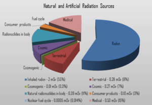
Natural and Artificial Radiation SourcesRadiation is all around us. In, around, and above the world we live in. It is a natural energy force that surrounds us. It is a part of our natural world that has been here since the birth of our planet. All living creatures, from the beginning of time, have been, and are still being, exposed to ionizing radiation. Ionizing radiation is generated through nuclear reactions, nuclear decay, by very high temperature, or via acceleration of charged particles in electromagnetic fields. But in general, there are two broad categories of radiation sources:

  • Natural Background Radiation. Natural background radiation includes radiation produced by the Sun, lightnings, primordial radioisotopes or supernova explosions etc.
  • Man-Made Sources of Radiation. Man-made sources include medical uses of radiation, residues from nuclear tests, industrial uses of radiation etc.

Special Reference: Sources and effects of ionizing radiation, Annex B. UNSCEAR. New York, 2010. ISBN: 978-92-1-142274-0.

Man-made Sources of Radiation

Since ionizing radiation has many industrial, and medical uses, people can be exposed also to man-made sources of radiation. Man-made sources include medical uses of radiation, residues from nuclear tests, industrial uses of radiation, television, and numerous other radiation producing devices. For example, in some kind of smoke detectors, you can meet man-made radionuclides such as americium-241. This man-made radionuclide is used to ionize air and to detect smoke.

It must be noted, most of these exposures are very low in intensity and the total dose and does not posse larger health effects. In each case, usefulness of ionizing radiation must be balanced with its hazards. Nowadays a compromise was found and most of uses of radiation are optimized. Today it is almost unbelievable that x-rays was, at one time, used to find the right pair of shoes (i.e. shoe-fitting fluoroscopy). Measurements made in recent years indicate that the doses to the feet were in the range 0.07 – 0.14 Gy for a 20 second exposure. This practice was halted when the risks of ionizing radiation were better understood.

There are two distinct groups exposed to man-made radiation sources. The United Nations Scientific Committee on the Effects of Atomic Radiation (UNSCEAR) itemized types of human exposures as:

  • public exposure, which is the exposure of individual members of the public and of the population in general
  • occupational radiation exposure, which is the exposure of workers in situations where their exposure is directly related to or required by their work

Public Exposure

In general, the following man-made sources expose the public to radiation:

  • Medical Exposures (by far, the most significant man-made source)
    • Diagnostic x-rays
    • Nuclear medicine procedures (iodine-131, cesium-137, technetium-99m etc.)
  • Consumer Products
    • Building and road construction materials
    • Smoking cigarettes (polonium-210)
    • Combustible fuels, including gas and coal
    • X-ray security systems
    • Televisions
    • Smoke detectors (americium)
    • Lantern mantles (thorium)

To a lesser degree, the public is also exposed to radiation from the nuclear fuel cycle, from uranium mining and milling to disposal of used (spent) fuel. Noteworthy, the public is also exposed to radiation from so called “enhanced sources of naturally occurring radioactive material”. This means also industries such as metal mining, coal mining and power production from coal creates additional exposures due to densification of naturally occurring radionuclides. The public receives some minimal exposure from the transportation of radioactive materials and fallout from nuclear weapons testing and reactor accidents (such as Chernobyl).

For that reason, most regulatory bodies require to limit the maximum radiation exposure to individual members of the public to 100 mrem (1 mSv) per year.

Occupational Exposure

As was written, occupational exposure is the exposure of workers in situations where their exposure is directly related to or required by their work. According to ICRP, occupational exposure refers to all exposure incurred by workers in the course of their work, with the exception of

  1. excluded exposures and exposures from exempt activities involving radiation or exempt sources
  2. any medical exposure
  3. the normal local natural background radiation.

In general, occupationally exposed individuals work in the following areas:

  • Fuel cycle facilities
  • Industrial radiography
  • Radiology departments (medical)
  • Nuclear medicine departments
  • Radiation oncology departments
  • Nuclear power plants
  • Government and university research laboratories

Such individuals are exposed to varying types and amounts of radiation, depending on their specific jobs and the sources with which they work. For that reason, most regulatory bodies require to limit occupational exposure to adults working with radioactive material to 5000 mrem (50 mSv) per year. Toward that end, employers carefully monitor the exposure of these individuals using instruments called dosimeters worn on a position of the body representative of its exposure. In most situations of occupational exposure the effective dose, E, can be derived from operational quantities using the following formula:
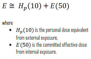
Occupational Exposure - External and Internal.

The committed dose is a dose quantity that measures the stochastic health risk due to an intake of radioactive material into the human body.

See also: Dose Monitoring

Medical Exposures – Doses from Medical Radiation Sources

Radiation is used in variety of medical examinations and treatments. Doses from medical radiation sources are naturally determined, whether a person underwent a treatment or not. In general, radiation exposures from medical diagnostic examinations are low (especially in diagnostic uses). Doses may be also high (only for therapeutic uses), but in each case, they must be always justified by the benefits of accurate diagnosis of possible disease conditions or by benefits of accurate treatment. These doses include contributions from medical and dental diagnostic radiology (diagnostic X-rays), clinical nuclear medicine and radiation therapy.

The medical use of ionizing radiation remains a rapidly changing field. In any case, usefulness of ionizing radiation must be balanced with its hazards. Nowadays a compromise was found and most of uses of radiation are optimized. Today it is almost unbelievable that x-rays was, at one time, used to find the right pair of shoes (i.e. shoe-fitting fluoroscopy). Measurements made in recent years indicate that the doses to the feet were in the range 0.07 – 0.14 Gy for a 20 second exposure. This practice was halted when the risks of ionizing radiation were better understood.

In the following points we try to express enormous ranges of radiation exposure as well as a few doses from medical sources.

  • 1 µSv – Eating one banana
  • 1 µSv – Extremity (hand, foot, etc.) X-ray
  • 5 µSv – Dental X-ray
  • 10 µSv – Average daily dose received from natural background
  • 40 µSv – A 5-hour airplane flight
  • 100 µSv – Chest X-ray
  • 600 µSv – mammogram
  • 1 000 µSv – Dose limit for individual members of the public, total effective dose per annum
  • 3 650 µSv – Average yearly dose received from natural background
  • 5 800 µSv – Chest CT scan
  • 10 000 µSv – Average yearly dose received from natural background in Ramsar, Iran
  • 20 000 µSv – single full-body CT scan
  • 80 000 µSv – The annual local dose to localized spots at the bifurcations of segmental bronchi in the lungs caused by smoking cigarettes (1.5 packs/day).
  • 175 000 µSv – Annual dose from natural radiation on a monazite beach near Guarapari, Brazil.
  • 5 000 000 µSv – Dose that kills a human with a 50% risk within 30 days (LD50/30), if the dose is received over a very short duration.

As can be seen, low-level doses are common for everyday life.

Tobacco – Smoking Cigarettes – Radiation Dose

In addition to chemical, nonradioactive carcinogens, tobacco and tobacco smoke contain small amounts of lead-210 and polonium-210 both of which are radioactive carcinogens. It must be emphasized, cigarettes and tobacco contain also polonium-210, originating from the decay products of radon, which stick to tobacco leaves. Polonium-210 emits a 5.3 MeV alpha particle, which provides most of equivalent dose. Due to decay of polonium-210, the annual local dose to localized spots at the bifurcations of segmental bronchi in the lungs caused by smoking cigarettes (1.5 packs/day) is about 80 mSv/year. Heavy smoking results in a dose of 160 mSv/year. This dose is not readily comparable to the radiation protection limits, since the latter deal with whole body doses, while the dose from smoking is delivered to a very small portion of the body. Many researchers believe that doses from polonium-210 are the origin of the high incidence of lung cancer among smokers.

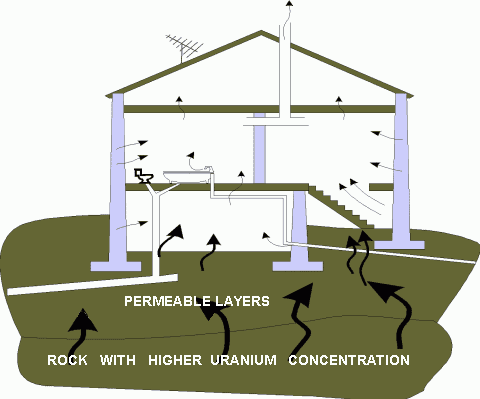
Recall, lead-210 and polonium-210 are daughter nuclei of radon-222. Radon-222 is a gas produced by the decay of radium-226. Both are a part of the natural uranium series. Since uranium is found in soil throughout the world in varying concentrations, also dose from gaseous radon is varying throughout the world. Radon-222 is the most important and most stable isotope of radon. It has a half-life of only 3.8 days, making radon one of the rarest elements since it decays away so quickly. An important source of natural radiation is radon gas, which seeps continuously from bedrock but can, because of its high density, accumulates at the ground. The fact radon is gas plays a crucial role in spreading of all its daughter nuclei. As radon-222 decays into lead-210, lead-210 can be attached to dust of moisture particles and it can be sticked to tobacco leaves. When these particles are concentrated by smoking and inhaled as smoke, some of lead-210 is retained by the body. Since lead-210 is a weak beta emitter, it does not cause major doses, but polonium-210 does.

See also: Radon – Health Effects

The polonium-210, the decay product of lead-210, emits a 5.3 MeV alpha particle, which provides most of equivalent dose. Alpha particles, that belongs to high-LET radiation, are fairly massive and carry a double positive charge, so they tend to travel only a short distance and do not penetrate very far into tissue if at all. However alpha particles will deposit their energy over a smaller volume (possibly only a few cells if they enter a body) and cause more damage to those few cells (more than 80 % of the absorbed energy from radon is due to the alpha particles). Therefore, the radiation weighting factor for alpha radiation is equal to 20. An absorbed dose of 1 mGy by alpha particles will lead to an equivalent dose of 20 mSv.

Special Reference: Sources and effects of ionizing radiation, Annex B. UNSCEAR. New York, 2010. ISBN: 978-92-1-142274-0.

Nuclear Fallout – Radiation Doses

In general, nuclear fallout is the residual radioactive material from a nuclear blast that “falls out” of the sky after an atmospheric explosion.  Fallout can also refer to nuclear reactor accidents, although a nuclear reactor does not explode like a nuclear weapon. The isotopic signature of fallout from nuclear blast is very different from the fallout from a serious power reactor accident.

In case of radiation doses from fallout, we consider the residual radioactive material from nuclear tests (not from reactor accidents) that were performed particularly in the two periods from 1954 to 1958 and from 1961 to 1962. According to the UNSCEAR, about 502 atmospheric tests, with a total fission and fusion yield of 440 Mt, were conducted.

Special Reference: Sources and effects of ionizing radiation, Annex B. UNSCEAR. New York, 2010. ISBN: 978-92-1-142274-0.

Fallout from a nuclear tests consist of fission fragments and neutron activation products. When a blast takes place on the ground or in the atmosphere near the ground, large amounts of activation products are formed also from surface materials. The fallout is particularly significant in the neighborhood of the test site, since the larger particles and most of debris lands on the ground (local fallout). But smaller particles  may remain aloft in the upper atmosphere for years. These particles can be therefore distributed nearly uniformly around the world, and contribute to so called global fallout. Equivalent doses from global fallout dropped from about 130 μSv/year in 1963 to about 10 μSv/year in recent years.

Radiation Exposures from Electricity Generation

In this chapter, we would like to discuss a very interesting fact. It is generally known, the increasing use of nuclear power and electricity generation using nuclear reactors will lead to a small but increasing radiation dose to the general public. But it is not generally known, power generation from coal also creates additional exposures, and, what is more interesting, while exposure levels are very low, the coal cycle contributes more than half of the total radiation dose to the global population from electricity generation. The nuclear fuel cycle contributes less than one-fifth of this. The collective dose, which are defined as the sum of all individual effective doses in a group of people over the time period or during the operation being considered due to ionizing radiation, is:

  • 670-1400 man Sv for coal cycle, depending on the age of the power plant,
  • 130 man Sv for nuclear fuel cycle,
  • 5-160 man Sv for geothermal power,
  • 55 man Sv for natural gas
  • 0.03 man Sv for oil

Yes, these results should be seen from the perspective of the share of each technology in worldwide electricity production. Since 40 per cent of the world’s energy was produced by the coal cycle in 2010, and 13 per cent by nuclear, the normalized collective dose will be about the same:

  • 0.7 – 1.4 man Sv/GW.a (man sievert per gigawatt year) for coal cycle
  • 0.43 man Sv/GW.a (man sievert per gigawatt year) for nuclear fuel cycle

Special Reference: Sources and effects of ionizing radiation, UNSCEAR 2016 – Annex B. New York, 2017. ISBN: 978-92-1-142316-7.

The above doses are related to public exposure. If we consider occupational exposure, regarding the mining of rare earth metals needed for construction, by far the largest collective dose to workers per unit of electricity generated assessed by the UNSCEAR came from solar power, followed by wind power. For solar power, occupational collective dose normalized to energy is a factor of forty and eighty larger than for the nuclear fuel cycle and coal cycle, respectively.

Note that, the collective effective dose is often used to estimate the total health effects, but according to the ICRP this should be avoided (see more: Collective Dose).

References:

Radiation Protection:

  1. Knoll, Glenn F., Radiation Detection and Measurement 4th Edition, Wiley, 8/2010. ISBN-13: 978-0470131480.
  2. Stabin, Michael G., Radiation Protection and Dosimetry: An Introduction to Health Physics, Springer, 10/2010. ISBN-13: 978-1441923912.
  3. Martin, James E., Physics for Radiation Protection 3rd Edition, Wiley-VCH, 4/2013. ISBN-13: 978-3527411764.
  4. U.S.NRC, NUCLEAR REACTOR CONCEPTS
  5. U.S. Department of Energy, Nuclear Physics and Reactor Theory. DOE Fundamentals Handbook, Volume 1 and 2. January 1993.

Nuclear and Reactor Physics:

  1. J. R. Lamarsh, Introduction to Nuclear Reactor Theory, 2nd ed., Addison-Wesley, Reading, MA (1983).
  2. J. R. Lamarsh, A. J. Baratta, Introduction to Nuclear Engineering, 3d ed., Prentice-Hall, 2001, ISBN: 0-201-82498-1.
  3. W. M. Stacey, Nuclear Reactor Physics, John Wiley & Sons, 2001, ISBN: 0- 471-39127-1.
  4. Glasstone, Sesonske. Nuclear Reactor Engineering: Reactor Systems Engineering, Springer; 4th edition, 1994, ISBN: 978-0412985317
  5. W.S.C. Williams. Nuclear and Particle Physics. Clarendon Press; 1 edition, 1991, ISBN: 978-0198520467
  6. G.R.Keepin. Physics of Nuclear Kinetics. Addison-Wesley Pub. Co; 1st edition, 1965
  7. Robert Reed Burn, Introduction to Nuclear Reactor Operation, 1988.
  8. U.S. Department of Energy, Nuclear Physics and Reactor Theory. DOE Fundamentals Handbook, Volume 1 and 2. January 1993.
  9. Paul Reuss, Neutron Physics. EDP Sciences, 2008. ISBN: 978-2759800414.

See also:

Sources of Radiation

We hope, this article, Man-made Sources of Radiation, helps you. If so, give us a like in the sidebar. Main purpose of this website is to help the public to learn some interesting and important information about materials and their properties.

What is Natural Background Radiation – Definition

Natural background radiation is ionizing radiation, that originates from a variety of natural sources. This radiation is not associated with any human activity. Material Properties

Natural and Artificial Radiation SourcesRadiation is all around us. In, around, and above the world we live in. It is a natural energy force that surrounds us. It is a part of our natural world that has been here since the birth of our planet. All living creatures, from the beginning of time, have been, and are still being, exposed to ionizing radiation. Ionizing radiation is generated through nuclear reactions, nuclear decay, by very high temperature, or via acceleration of charged particles in electromagnetic fields.

Natural Background Radiation

Natural background radiation is ionizing radiation, that originates from a variety of natural sources. All living creatures, from the beginning of time, have been, and are still being, exposed to ionizing radiation. This radiation is not associated with any human activity.  There are radioactive isotopes in our bodies, houses, air, water and in the soil. We all are also exposed to radiation from outer space.

Sources of Natural Background Radiation

We divide all these natural radiation sources into three groups:

Cosmic Radiation

Cosmic Radiation - Natural Source of Radiation
Source: nasa.gov License: Public Domain

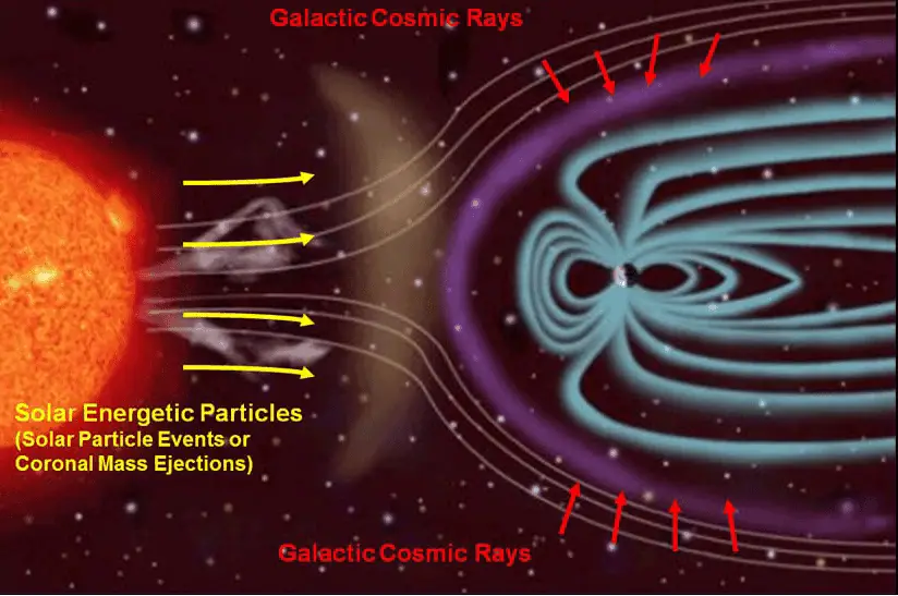
Cosmic radiation refers to sources of radiation in the form of cosmic rays that come from the sun or from outer space. At ground level the muons, with energies mostly between 1 and 20 GeV, contribute about 75 % of the absorbed dose rate in free air. The remainder comes from electrons produced by the muons or present in the electromagnetic cascade. The annual cosmic ray dose at sea level is around 0.27 mSv (27 mrem). If you live at higher elevations or are a frequent airline passenger, this exposure can be significantly higher, since the atmosphere is thinner here. The effects of the earth’s magnetic field also determines the dose from cosmic radiation.

Cosmic radiation can be divided into different types according to its origin. There are three main sources of such radiation:

  • Solar Cosmic Radiation. Solar cosmic radiation refers to sources of radiation in the form of high-energy particles (predominantly protons) emitted by the sun, primarily in solar particle events (SPEs).
  • Galactic Cosmic Radiation. Galactic cosmic radiation, GCR, refers to sources of radiation in the form of high-energy particles originating outside the solar system, but generally from within our Milky Way galaxy.
  • Radiation from Earth’s Radiation Belts (van Allen belts). Van Allen radiation belts are zones of high-energy particles (especially protons) trapped by earth’s magnetic field.

Natural Background in Airplane – Radiation in Flight

Exposure to cosmic radiation increases rapidly with altitude. In flight there are two principal sources of natural radiation to consider: Galactic Cosmic Rays which are always present, and Solar Proton Events, sometimes called Solar Cosmic Ray (SCR) events, which occur sporadically. The dose rate from cosmic radiation varies in different parts of the world and it depends strongly on the geomagnetic field, altitude, and solar cycle. The radiation field at aircraft altitudes consist of neutrons, protons and pions. In flight, neutrons contribute 40 – 80% of the equivalent dose, depending on the geomagnetic field, altitude, and solar cycle. The cosmic radiation dose rate on airplanes is so high (but not dangerous) that, according to the United Nations UNSCEAR 2000 Report, airline flight crew workers receive more dose on average than any other worker, including those in nuclear power plants.

The ground level dose rate is on average about 0.10 μSv/h, but at the maximum flight altitude (8.8 km or 29,000 ft) it can reach about 2.0 μSv/h (or even higher values). A dose rate of 4 μSv/h may be used to represent the average dose rate for all long haul flights (due to higher altitudes). It must be added, for supersonic planes like the Concorde, that could make a transatlantic flight in 3.5 hours, the exposure rate (about 9 μSv/h) at their altitude of 18 km was increased enough to result in the same cosmic ray exposure per crossing as for conventional jets trundling along at about 8 km.

Earth’s Magnetic Field as Radiation Shield

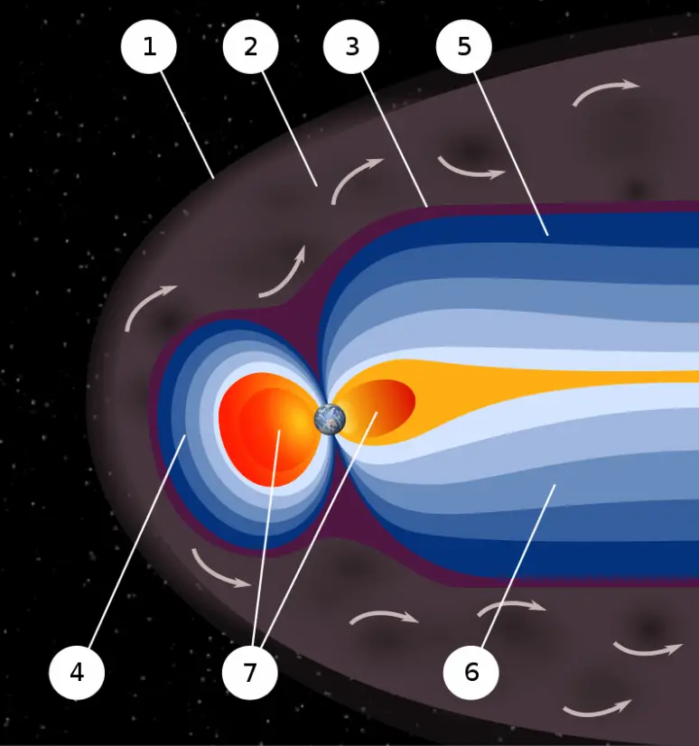
magnetosphere - earth magnetic field
An artist’s rendering of the structure of a magnetosphere: 1) Bow shock. 2) Magnetosheath. 3) Magnetopause. 4) Magnetosphere. 5) Northern tail lobe. 6) Southern tail lobe. 7) Plasmasphere. Source: nasa.gov License: Public Domain

Earth’s magnetic field provides a vital radiation shield of cosmic radiation. In addition to a protective atmosphere, we are also lucky that Earth has a magnetic field. Magnetic field extends several tens of thousands of kilometers into space, protecting the Earth from the charged particles of the solar wind and cosmic rays that would otherwise strip away the upper atmosphere, including the ozone layer that protects the Earth from harmful ultraviolet radiation. It shields us from the full effects of the solar wind and GCR. Without this protection, Earth’s biosphere might not exist as it does today, or would be at least limited to the subsurface.  Earth’s magnetic field provides also a radiation shield for astronauts and the ISS itself, because it is in low Earth orbit.

Calculations of the loss of carbon dioxide from the atmosphere of Mars, resulting from scavenging of ions by the solar wind, indicate that the dissipation of the magnetic field of Mars caused a near total loss of its atmosphere.

Terrestrial Radiation

Terrestrial radiation refers to sources of radiation that are in the soil, water, and vegetation. The major isotopes of concern for terrestrial radiation are uranium and the decay products of uranium, such as thorium, radium, and radon. The average dose rate that originates from terrestrial nuclides (except radon exposure) is about 0.057 µGy/hr. The maximum values have been measured on monazite sand in Guarapari, Brazil (up to 50 µGy/hr and in Kerala, India (about 2 µGy/hr), and on rocks with a high radium concentration in Ramsar, Iran (from 1 to 10 µGy/hr).

radon - mitigation - house
The radon gas can penetrate into the house through cracks (due to a chimney effect) in the floor and walls of the basement. Source: suro.cz

The average annual radiation dose to a person from radon is about 2 mSv/year and it may vary over many orders of magnitude from place to place. Radon is so important, that it is usually treated separately. Radon is a colorless, odorless, tasteless noble gas, which seeps continuously from bedrock but can, because of its high density, accumulate in poorly ventilated houses. The fact radon is gas plays a crucial role in spreading of all its daughter nuclei. Simply radon is a transport medium from bedrock to atmosphere (or inside buildings) for its short-lived decay products (Pb-210 and Po-210), that posses much more health risks.

Internal Radiation

In addition to the cosmic and terrestrial sources, all people also have radioactive potassium-40, carbon-14, lead-210, and other isotopes inside their bodies from birth.

These isotopes are especially potassium-40, carbon-14 and also the isotopes of uranium and thorium. The variation in radiation dose from one person to another is not as great as the variation in dose from cosmic and terrestrial sources. The average annual radiation dose to a person from internal radioactive materials other than radon is about 0.3 mSv/year of which:

  • 0.2 mSv/year comes from potassium-40,
  • 0.12 mSv/year comes from the uranium and thorium series,
  • 12 μSv/year comes from carbon-40.

Background Radiation and Health Hazard

You can not go through life without radiation. The danger of ionizing radiation lies in the fact that the radiation is invisible and not directly detectable by human senses. People can neither see nor feel radiation, yet it deposits energy to the molecules of the body.

LNT Model and Hormesis Model
Alternative assumptions for the extrapolation of the cancer risk vs. radiation dose to low-dose levels, given a known risk at a high dose: LNT model, and hormesis model.

But don’t worry, the doses from background radiation are usually very small (except radon exposure). Low dose here means additional small doses comparable to the normal background radiation (10 µSv = average daily dose received from natural background). The problem is that, at very low doses, it is practically impossible to correlate any irradiation with certain biological effects. This is because the baseline cancer rate is already very high and the risk of developing cancer fluctuates 40% because of individual life style and environmental effects, obscuring the subtle effects of low-level radiation.

Secondly, and this is crucial, the truth about low-dose radiation health effects still needs to be found. It is not exactly known, whether these low doses of radiation are detrimental or beneficial (and where is the threshold).  Government and regulatory bodies assume a LNT model instead of a threshold or hormesis not because it is the more scientifically convincing, but because it is the more conservative estimate. Problem of this model is that it neglects a number of defence biological processes that may be crucial at low doses. The research during the last two decades is very interesting and show that small doses of radiation given at a low dose rate stimulate the defense mechanisms. Therefore the LNT model is not universally accepted with some proposing an adaptive dose–response relationship where low doses are protective and high doses are detrimental. Many studies have contradicted the LNT model and many of these have shown adaptive response to low dose radiation resulting in reduced mutations and cancers. This phenomenon is known as radiation hormesis.

According to the radiation hormesis hypothesis, radiation exposure comparable to and just above the natural background level of radiation is not harmful but beneficial, while accepting that much higher levels of radiation are hazardous. Arguments for hormesis center around some large-scale epidemiological studies and the evidence from animal irradiation experiments, but most notably the recent advances in knowledge of the adaptive response. Proponents of radiation hormesis typically claim that radio-protective responses in cells and the immune system not only counter the harmful effects of radiation but additionally act to inhibit spontaneous cancer not related to radiation exposure.

See also: LNT Model

References:

Radiation Protection:

  1. Knoll, Glenn F., Radiation Detection and Measurement 4th Edition, Wiley, 8/2010. ISBN-13: 978-0470131480.
  2. Stabin, Michael G., Radiation Protection and Dosimetry: An Introduction to Health Physics, Springer, 10/2010. ISBN-13: 978-1441923912.
  3. Martin, James E., Physics for Radiation Protection 3rd Edition, Wiley-VCH, 4/2013. ISBN-13: 978-3527411764.
  4. U.S.NRC, NUCLEAR REACTOR CONCEPTS
  5. U.S. Department of Energy, Nuclear Physics and Reactor Theory. DOE Fundamentals Handbook, Volume 1 and 2. January 1993.

Nuclear and Reactor Physics:

  1. J. R. Lamarsh, Introduction to Nuclear Reactor Theory, 2nd ed., Addison-Wesley, Reading, MA (1983).
  2. J. R. Lamarsh, A. J. Baratta, Introduction to Nuclear Engineering, 3d ed., Prentice-Hall, 2001, ISBN: 0-201-82498-1.
  3. W. M. Stacey, Nuclear Reactor Physics, John Wiley & Sons, 2001, ISBN: 0- 471-39127-1.
  4. Glasstone, Sesonske. Nuclear Reactor Engineering: Reactor Systems Engineering, Springer; 4th edition, 1994, ISBN: 978-0412985317
  5. W.S.C. Williams. Nuclear and Particle Physics. Clarendon Press; 1 edition, 1991, ISBN: 978-0198520467
  6. G.R.Keepin. Physics of Nuclear Kinetics. Addison-Wesley Pub. Co; 1st edition, 1965
  7. Robert Reed Burn, Introduction to Nuclear Reactor Operation, 1988.
  8. U.S. Department of Energy, Nuclear Physics and Reactor Theory. DOE Fundamentals Handbook, Volume 1 and 2. January 1993.
  9. Paul Reuss, Neutron Physics. EDP Sciences, 2008. ISBN: 978-2759800414.

See also:

Sources of Radiation

We hope, this article, Natural Background Radiation, helps you. If so, give us a like in the sidebar. Main purpose of this website is to help the public to learn some interesting and important information about materials and their properties.

What is Carbon-14 – Production – Properties – Decay – Definition

Carbon-14 is a naturally occurring isotope. Radioactive carbon-14 has a half-life of 5730 years and undergoes β− decay. Carbon-14 is continuously formed in the upper atmosphere

Carbon-14

The only cosmogenic radionuclide to make a significant contribution to internal exposure of human is carbon-14. Radioactive carbon-14 has a half-life of 5730 years and undergoes β− decay, where the neutron is converted into a proton, an electron, and an electron antineutrino:

beta decay
Beta decay of C-14 nucleus.

In spite of this short half-life compared to the age of the earth, carbon-14 is a naturally occurring isotope. Its presence can be explained by the following simple observation. Our atmosphere contains many gases, including nitrogen-14. Besides, the atmosphere is constantly bombarded with high energy cosmic rays, consisting of protons, heavier nuclei, or gamma rays. These cosmic rays interact with nuclei in the atmosphere, and produce also high-energy neutrons. These neutrons produced in these collisions can be absorbed by nitrogen-14 to produce an isotope of carbon-14:

carbon-14 dating - formation

Carbon-14 can also be produced in the atmosphere by other neutron reactions, including in particular 13C(n,γ)14C and 17O(n,α)14C. As a result, carbon-14 is continuously formed in the upper atmosphere by the interaction of cosmic rays with atmospheric nitrogen. On average just one out of every 1.3 x 10¹² carbon atoms in the atmosphere is a radioactive carbon-14 atom. As a result, all living biological substances contain the same amount of C-14 per gram of carbon, that is 0.3 Bq of carbon-14 activity per gram of carbon. Carbon-14 is present in the human body (13kg of carbon in 70kg human) at a level of about 3700 Bq (0.1 μCi) with a biological half-life of 40 days. Note that, biological half-life is the time taken for the amount of a particular element in the body to decrease to half of its initial value due to elimination by biological processes alone. However, a carbon atom is in the genetic information of about half the cells, while potassium is not a component of DNA. The decay of a carbon-14 atom inside DNA in one person happens about 50 times per second, changing a carbon atom to one of nitrogen.

The annual dose from carbon-14 is estimated to be about 12 μSv/year.

As long as the biological system is alive the level is constant due to constant intake of all isotopes of carbon. When the biological system dies, it stops exchanging carbon with its environment, and from that point onwards the amount of carbon-14 it contains begins to decrease as the carbon-14 undergoes radioactive decay.

See also: Carbon-14 Dating

References:

Radiation Protection:

  1. Knoll, Glenn F., Radiation Detection and Measurement 4th Edition, Wiley, 8/2010. ISBN-13: 978-0470131480.
  2. Stabin, Michael G., Radiation Protection and Dosimetry: An Introduction to Health Physics, Springer, 10/2010. ISBN-13: 978-1441923912.
  3. Martin, James E., Physics for Radiation Protection 3rd Edition, Wiley-VCH, 4/2013. ISBN-13: 978-3527411764.
  4. U.S.NRC, NUCLEAR REACTOR CONCEPTS
  5. U.S. Department of Energy, Nuclear Physics and Reactor Theory. DOE Fundamentals Handbook, Volume 1 and 2. January 1993.

Nuclear and Reactor Physics:

  1. J. R. Lamarsh, Introduction to Nuclear Reactor Theory, 2nd ed., Addison-Wesley, Reading, MA (1983).
  2. J. R. Lamarsh, A. J. Baratta, Introduction to Nuclear Engineering, 3d ed., Prentice-Hall, 2001, ISBN: 0-201-82498-1.
  3. W. M. Stacey, Nuclear Reactor Physics, John Wiley & Sons, 2001, ISBN: 0- 471-39127-1.
  4. Glasstone, Sesonske. Nuclear Reactor Engineering: Reactor Systems Engineering, Springer; 4th edition, 1994, ISBN: 978-0412985317
  5. W.S.C. Williams. Nuclear and Particle Physics. Clarendon Press; 1 edition, 1991, ISBN: 978-0198520467
  6. G.R.Keepin. Physics of Nuclear Kinetics. Addison-Wesley Pub. Co; 1st edition, 1965
  7. Robert Reed Burn, Introduction to Nuclear Reactor Operation, 1988.
  8. U.S. Department of Energy, Nuclear Physics and Reactor Theory. DOE Fundamentals Handbook, Volume 1 and 2. January 1993.
  9. Paul Reuss, Neutron Physics. EDP Sciences, 2008. ISBN: 978-2759800414.

See also:

Cosmogenic Radionuclides


We hope, this article, Carbon-14 – Production – Properties – Decay, helps you. If so, give us a like in the sidebar. Main purpose of this website is to help the public to learn some interesting and important information about materials and their properties.