The variety of techniques available can also be divided into two groups: surface methods, which are used to identify surface and near surface defects such as cracks and surface porosity, and subsurface methods, that can be used to detect defects that lie under the material’s surface.
- Surface methods
- Subsurface methods
Ultrasonic Testing
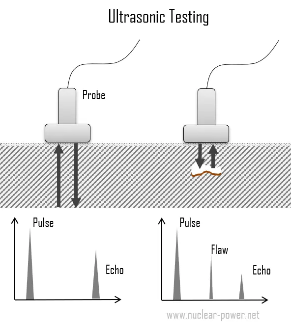 Ultrasonic testing is a broad group of NDT techniques based on the propagation of ultrasonic waves in the object or material tested. The most commonly used ultrasonic testing technique is pulse echo, wherein high-frequency sound wave beams (normally ranging from 1-5 MHz) is introduced into a test object and reflections (echoes) are returned to a receiver from internal imperfections or from the part’s geometrical surfaces.
The basic method of ultrasonic testing is transforming a voltage pulse to an ultrasonic pulse using a transducer. Transducers used for traditional UT consist of a piezoelectric crystal enclosed within a plastic or stainless steel housing. The piezoelectric crystals expand when electrically charged, thus generating an acoustic wave. The signal travels through the object with respect to its geometry and existing defects and then is either transmitted to another transducer or reflected back to the original transducer. Defects are detected if they produce a change in the acoustic impedance in the path of the ultrasonic beam. An open crack filled with air has very low acoustic impedance so it reflects virtually all the acoustic energy incident on it. Hence, the sound waves travel through the material and are reflected back from cracks or flaws. Defects and flaws affect its way and a small portion of the pulse will be sent back to the transducer/receiver before it hits the end of the object.
Since the speed of sound in the parent material is known, the reflected sound energy is then displayed versus time and analyzed to define the presence and location of flaws or discontinuities.
Advantages and Disadvantages of Ultrasonic Testing
The advantages and disadvantages of ultrasonic testing method are as follows:
Advantages:
- UT is flexible, portable and has high penetration depth.
- High sensitivity, permitting the detection of extremely small flaws.
- It is applicable in wide range of industries
- Some capability of estimating the size, orientation, shape and nature of defects.
- Unlike radiographic testing, there is no health or environmental risk involved
Disadvantages:
- It requires highly-trained operators and requires careful attention by experienced technicians.
- Unable or not efficient in detecting planar defect that are in parallel with the direction of sound wave
- Parts that are rough, irregular in shape, very small or thin, or not homogeneous are difficult to inspect.
- Can be very expensive
Radiographic Testing
Radiographic testing (RT) involves the use of penetrating gamma or X-radiation to examine parts and products for imperfections. It is one of the conventional NDT methods which has been in use over decades and is still being used by companies around the world.
- X-rays, also known as X-radiation, refers to electromagnetic radiation (no rest mass, no charge) of high energies. Most X-rays have a wavelength ranging from 0.01 to 10 nanometers (3×10¹⁶ Hz to 3×10¹⁹ Hz), corresponding to energies in the range 100 eV to 100 keV. X-ray wavelengths are shorter than those of UV rays and typically longer than those of gamma rays. The distinction between X-rays and gamma rays is not so simple and has changed in recent decades. According to the currently valid definition, X-rays are emitted by electrons outside the nucleus, while gamma rays are emitted by the nucleus. X-rays can be generated by an X-ray tube, a vacuum tube that uses a high voltage to accelerate the electrons released by a hot cathode to a high velocity. Upon striking the target, the accelerated electrons are abruptly stopped and X-rays and heat are generated.
- Gamma rays, also known as gamma radiation, refers to electromagnetic radiation (no rest mass, no charge) of a very high energies. Since the gamma rays are in substance only a very high-energy photons, they are very penetrating matter and are thus biologically hazardous. Gamma rays can travel thousands of feet in air and can easily pass through the human body. Gamma rays are emitted by unstable nuclei in their transition from a high energy state to a lower state known as gamma decay. In most practical laboratory sources, the excited nuclear states are created in the decay of a parent radionuclide, therefore a gamma decay typically accompanies other forms of decay, such as alpha or beta decay.
In general, RT is method of inspecting materials for hidden subsurface defects by using the ability of X-rays or gamma rays to penetrate various materials of various thickness. The intensity of the radiation that penetrates and passes through the material is either captured by:
- a radiation sensitive film (Film Radiography)
- a planer array of radiation sensitive sensors (Real-time Radiography).
Principle of Operation
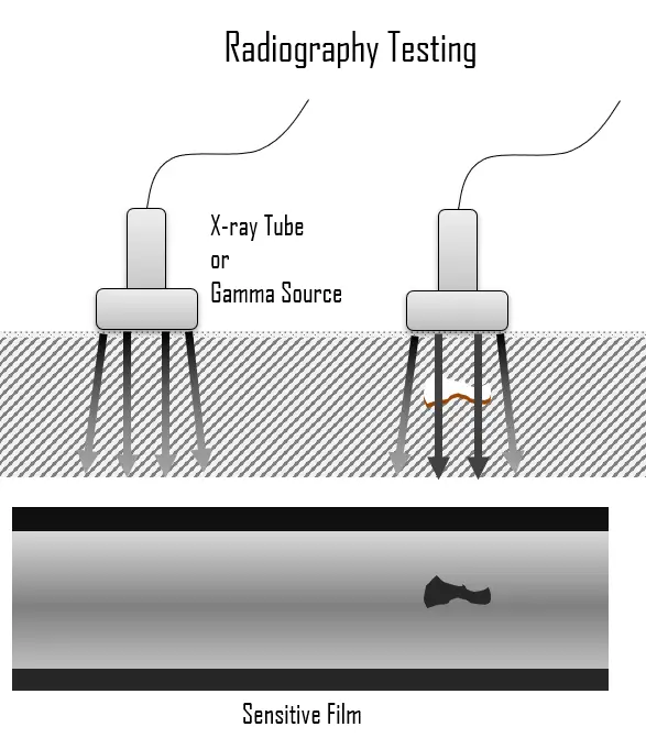 The radiation source can either be an X-ray machine or a radioactive source (Ir-192, Co-60, or in rare cases Cs-137). The choice between X-rays and gamma radiation depends on some factors such as thickness, contrast level and etc. For example X-rays typically work with lower amount of energy than gamma rays. The thickness is another parameter which influences the results. For example, at thicknesses more than 50 mm, the use of gamma rays increases significantly.
Radiation is directed through a part and onto film or other imaging media. The resulting radiograph shows the dimensional features of the part. Both in X-rays and gamma radiation as radiation passes more through the material the darker the film becomes on the image produced and, on the contrary, the more the ray is absorbed by the material the lighter is the image in those spots. Therefore, possible imperfections are indicated as density changes on the film in the same manner as a medical X-ray shows broken bones.
Radiographic testing is commonly used for weld verification in various industrial applications. In manufacturing, welds are commonly used to join two or more metal parts. The effects of welding on the material surrounding the weld can be detrimental —depending on the materials used and the heat input of the welding process used, the HAZ can be of varying size and strength. For example, the base metal must reach a certain temperature during the welding process, must cool at a specific rate, and must be welded with compatible materials or the joint may not be strong enough to hold the parts together, or cracks may form in the weld causing it to fail. Defects usually encountered include incomplete penetration, incomplete fusion, undercutting, porosity, and longitudinal cracking. These defects could cause a structure to break or a pipeline to rupture. Welds may be tested using NDT techniques such as industrial radiography or industrial CT scanning using X-rays or gamma rays, ultrasonic testing, liquid penetrant testing, magnetic particle inspection or via eddy current.
Advantages and Disadvantages
Advantages:
- It has a very few material limitations.
- Detection of internal defects for thick materials (e.g. pipelines).
- Minimal or no part preparation is required.
- One of the major advantages of RT is its documentation capability. RT provides images of the object under inspection.
- The probability of misinterpretation of results is minimized since each image can be reviewed by multiple operators.
Disadvantages:
- The impact of radiation to health and environment can be considered as one of major disadvantages of radiographic testing since a few seconds of being exposed to radiation can result in sever injuries.
- High degree of skill and experience is required for exposure and interpretation.
- The high voltage needed to create X-rays is dangerous for human health also.
- It is quite expensive method.
- Ineffective for planar defects and for surface defects.
We hope, this article, Subsurface NDT Methods, helps you. If so, give us a like in the sidebar. Main purpose of this website is to help the public to learn some interesting and important information about materials and their properties.